Touch : Spinal Pathways |
|
The sense of touch involves sensitive sensory receptors in the skin. Two types of sensory receptors respond to skin indentation (Merkel cell receptors; Type I slowly adapting mechanoreceptors) and lateral stretch of the skin (Ruffini end organs; Type II slowly adapting mechanoreceptors). Movement of objects over the skin are sensed by Meissner's Corpuscles, Pacinican Corpuscels and hair receptors. Meissner's corpuscles also respond to vibration with an optimum frequence of around 30 Hz, whereas the highly sensitive Pacinian Corpuscles respond optimally to vibrations of around 250Hz. Sometimes the perception of vibratory stimuli is divided into two types on the basis of optimal frequency: frequencies of ~30Hz are referred to as 'flutter', and frequencies of ~200-300 are referred to as 'vibration'. Vibration can also be sensed by muscle spindles. All of these sensory receptors have myelinated axons. The highest density of cutaneous receptors involved in touch is found in areas in which two point discrimination is greatest, such as the fingertips and lips. Two point discrimination can be measured by asking a blind-folded subject whether they feel one or two objects when the two points of a compass are applied to the skin; as the two points of the compass are brought closer together, the subject cannot distinguish the two points as being separate. |

Different groups of sensory receptors have axons with different diameters Proprioceptors from muscles and tendons, and touch receptors from the skin have large diameter axons. Thermoreceptors and Nociceptors have either small finely myelinated (Ad, A-delta) or unmyelinated axons (C fibres)
|
| Dorsal Columns Top |
|
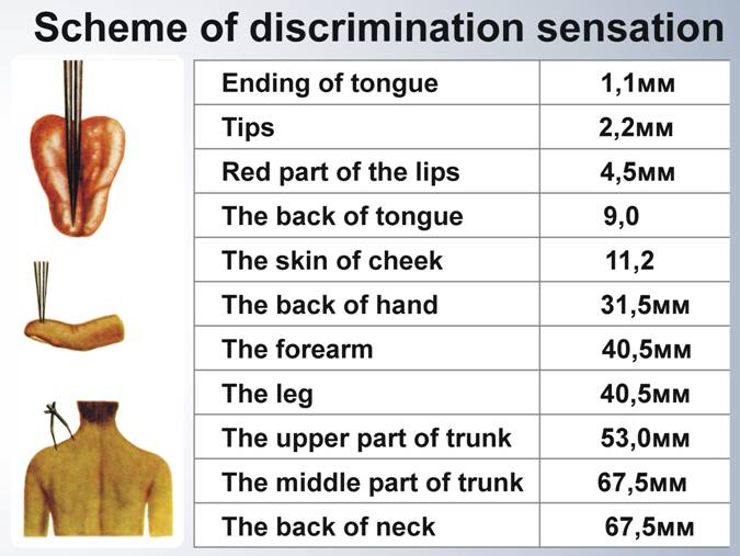
The dorsal columns contain the axon collaterals of primary sensory neurones and project rostrally to the dorsal column nuclei at the caudal end of the medulla. These sensory neurones have large diameter axons and are concerned with the senses of touch and vibration. In lower segments of the cord the dorsal columns are thin, and these fibres remain in a medial position. Axons from higher segments of the cord are added to the lateral edge of the dorsal columns. In the cervical region there are two distinct components of the doral columns - the fasciculus gracilis (medial) and fasciculus cuneatus (lateral). This pathways is specialised for sensory discrimination, and this can be examined using the two-point discrimination test. Subjects have to decide if a stimulus consiting of two points touching the skin is felt as one or two points. |
It can be seen from the table that the most sensitive discrimination occurs in the tongue and the finger tips. |
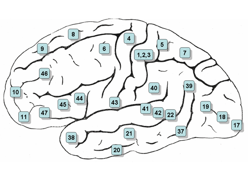
 * *The representation of the body surface on the primary somatosensory cortex, S1, (left) shows that areas with highest two-point discrimination have the greatest areas on the cortical map. Somatotopic Maps are found not only in S1, but also in the thalamus and dorsal column nuclei. |
|
| Dorsal Column Nuclei Top |
|
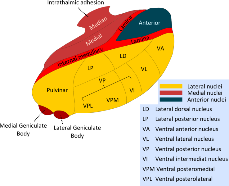
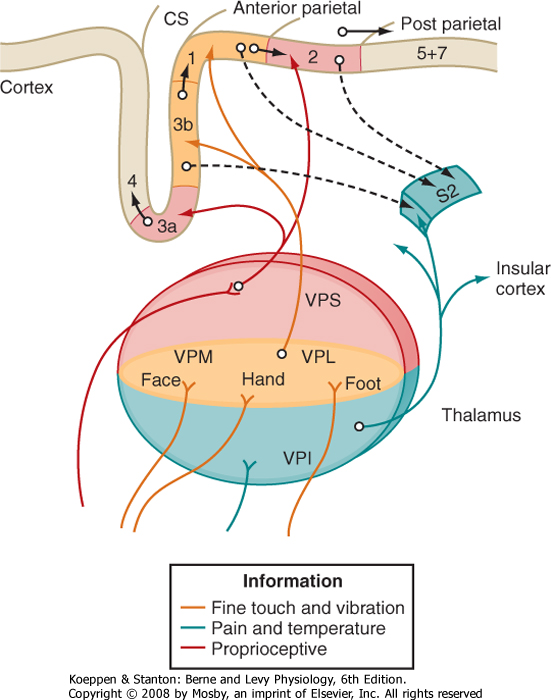
Dorsal Columns and Dorsal Column Nuclei. The neurones of the dorsal column nuclei are second order neurones, i.e. they are the second neurones in the chain of neurones carrying infomation to higher levels. They have axons that project to the ventro-posterior nuclei of the opposite side of the thalamus. These axons cross the midline within the medulla, and form the medial lemniscus, which passes through the pons and the dorsal tegmentum of the midbrain to reach the thalamus. In the dorsal columns, the afferent fibres from the lower limb are found medial to those from the upper limb. In the medial lemniscus at the level of the midbrain, the orietation is reversed, with the fibres from the upper limb being medial. Somatosensory fibers are arranged in such a way as to preserve spatial information in their position, forming a map of the body surface known as a somatotopic map. Somatotopic maps are found in the dorsal column nuclei, the medial lemniscus, the ventrobasal complex of the thalamus and the somatosensory cortex. There is a precise point-to-point pattern of projections from the body surface at each level in this pathway. Relay neurones maintain a high degree of modality specificity, i.e. vibration receptors, proprioceptors, and the various types of cutaneous receptors all preserve their private pathways through the dorsal column nuclei - there is no mixing of modalities within the different parts of the dorsal column nuclei. |
|
| |
|
Sensory processing in the dorsal column nuclei. Within the nucleus gracilis there is some sensory processing, known as lateral inhibition (surround inhibition), that involves inhibitory interneurones present within the dorsal column nuclei. The inhibition produced by these interneurones is arranged in a spatial pattern across the nucleus, so as to suppress the activity of projection neurones around the most active ones, thus emphasising the contrast between the most active neurons and the surrounding ones. This is also a feature of the visual pathway in the retina, lateral geniculate nucleus and visual cortex. The top half of the diagram indicates how impusles form the skin would be transmitter to relay neurones (projecting in the medial lemniscus) if there were no interneurones in the dorsal column nuclei. Discrimination of two points as separate entitites woud be difficult.
The bottom half of the diagram shows the role of inhibitory interneurones in the dorsal column nuclei (and thalamus). By inhibiting the surrounding neurones the site of the stimulus is clearer. Lateral inhibition or Surround Inhibition is a means of highlighting the activity of groups of active neurones by inhibiting the activity of neurones lateral to the centre of activity within the spatial map of the skin surface. This enhances the contrast between active and inactive areas.  * *
If a tactile stimulus is applied to a point on the fingertip, the primary afferents that discharge excite a group of neurones in the nucleus cuneatus; lateral inhibition causes cuneate neurones that carry information from areas of the fingertip lateral to the stimulus to reduce their activity. This mechanism, within the population of cuneate nucleus signalling tactile stimulation of the fingertip, allows the spatial map of the fingertip to emphasise the activity between the active sensory neurones and the rest, which are inhibited; in other words it helps to define the edge of the stimulated area. Note that afferents and interneurones are involved in post-synaptic and pre-synaptic inhibitory processes that provides the spatial distribution recognised at lateral inhibition.. One other feature of the diagram is the presence of descending pathways from the forebrain that can enhance the process of lateral inhibition. Lateral inhibition is a common mechanism within sensory systems, and is found in second and third order neurones in the somatosensory and visual pathways. It provides contrast enhancement which helps the brain to identify the edges of a tactile or visual stimulus. |
|